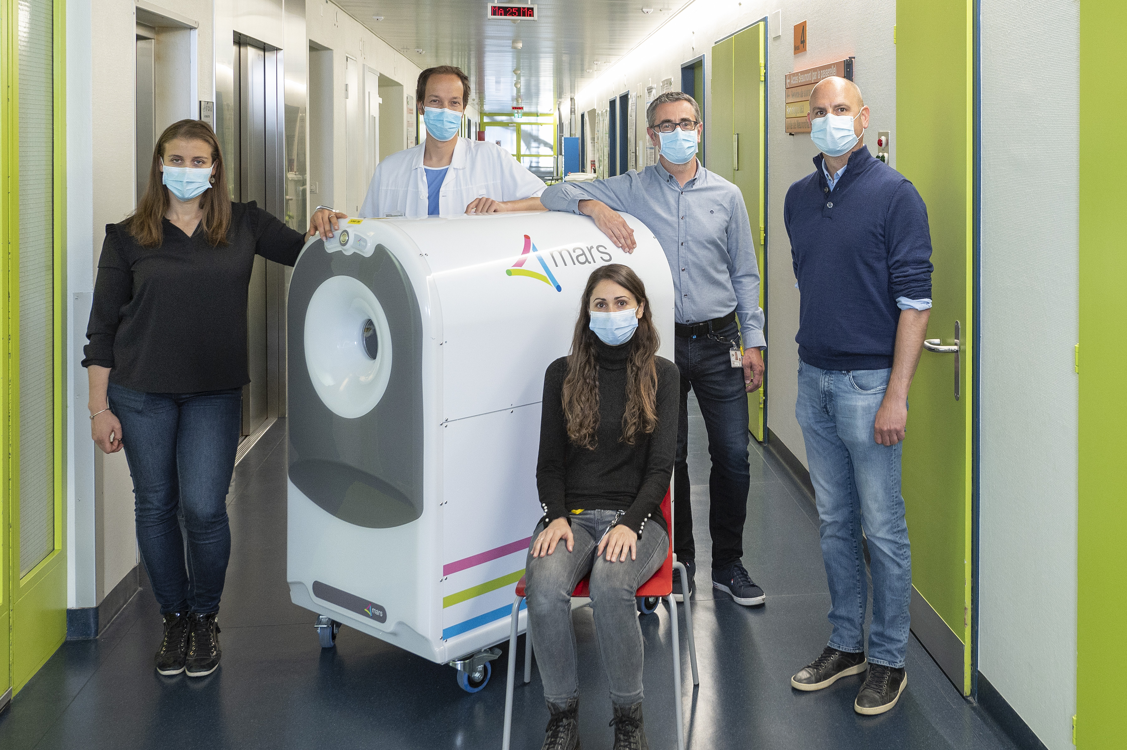
After years of development in New Zealand, MARS Bioimaging's 3D colour X-ray scanner has arrived in Europe, at Lausanne University Hospital (CHUV) in Switzerland. This event marks the first step towards the European part of the international clinical trials undertaken by MARS Bioimaging, a New Zealand company, to obtain the certifications allowing its medical use.
With the potential to monitor bone healing following a fracture, MARS Bioimaging’s scanner allows high resolution imaging around metal implants and can distinguish many different types of tissue without any use of contrast agents. Compared to existing technologies, images of such precision would enable significant progress to be made in diagnosing hand and wrist fractures and monitoring the healing process.
The entire team of radiologists and medical physicists at CHUV is eager to start the clinical use of the scanner. “The MARS scanner will allow us to improve our understanding of arthritis: how it develops and how to diagnose it. It should also help us develop the targeted therapies we are currently lacking for calcium crystal deposition diseases” explains Dr. Fabio Becce, Associate Physician and Senior Lecturer at Lausanne University Hospital (CHUV). “In practical terms, the MARS scanner gives us a unique opportunity to better identify and quantify urate and calcium crystal deposits in and around joints”.
“Reaching this incredible milestone is really exciting for us and our collaborators” says Professor Anthony Butler, President of MARS Bioimaging.
Trial of this technology in a Swiss hospital clearly demonstrates the pathway from experiments performed in a physics research laboratory to making a difference to patient health care
Professor Anthony Butler, President of MARS Bioimaging.
Since 2008, CERN and the New Zealand company have teamed up to develop a 3D colour X-ray scanner based on the Medipix3 technology, developed by the Medipix3 collaboration hosted by CERN. Inspired by particle physics detectors, Medipix3 and Timepix3 chips are now used for medical applications, in space, and for art authentication.
“In June 2021, CERN and MARS Bioimaging extended their current contract by five years, thus supporting MARS Bioimaging on their way to the US Food and Drug Administration and EU approval.”, explains Aurélie Pezous, from CERN’s Knowledge Transfer group. “The partnership between CERN, the Medipix3 Collaboration, and MARS Bioimaging shows how teaming up with health professionals is critical to enhance medical innovation.”
Beyond knowledge transfer, this cooperation highlights the potential of CERN Alumni. Various CERN Alumni have been involved in ensuring the scanner’s radiological safety. Amongst them is Lucia Gallego Manzano, former CERN fellow in radioprotection, now with the Institut de radiophysique (IRA) of CHUV.